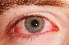
| Corneal Ulcers |
 |
|
|
| |
 |
What is it? |
| |
It is a bilateral conical protrusion of the central part of the cornea with thinning
of its central and inferior paracentral areas. Keratoconus is a degenerative disorder of the
eye in which structural changes within the cornea cause it to thin and change to a more
conical shape than its normal gradual curve. |
 |
What Causes it? |
| |
Often the cause of keratoconus is unknown. Some studies have found that keratoconus
runs in families, and that it happens more often in people with certain medical problems, including
certain allergic conditions. Some think that chronic eye rubbing can cause keratoconus. But most
often, there is no eye injury or disease that could explain why the eye starts to change.
Keratoconus usually begins in the teenage years, but it can also start in childhood or up to about
age 30. The changes in the shape of the cornea usually occur slowly over several years.
Someone with keratoconus will notice that vision slowly becomes distorted. The change can stop
at any time, or it can continue for several years. In most people who have keratoconus, both eyes
are eventually affected, although not always to the same extent. |
 |
Who are at risk? |
| |
These factors can increase your chances of developing keratoconus: Certain
diseases - The risk of developing keratoconus is higher if you have certain inherited
diseases or genetic conditions, such as Down syndrome. Keratoconus also is more likely
in people with allergies or asthma, connective tissue disorders and some retinal diseases.
Family history of keratoconus. |
 |
What are the symptoms & signs? |
| |
Symptoms: Initial symptom is impaired vision (in one eye it is more than the
fellow eye) due to irregular myopic astigmatism. This visual loss can only be improved
by hard contact lenses. Later on there is further impairment of vision and contact lens can
no longer correct the visual loss.
Signs:
1. Irregular retinoscopic reflex.
2. Distortion of mires of placido’s disc or of keratometer.
3. Vertical folds at the level of deep stroma and descemet,s membrane (Vogt’s sign).
4. Prominent corneal nerves.
5. Thinning of the central cornea with protrusion just below and nasal to the centre.
6. Munson,s sign – A ‘bulging’ or ‘tenting’ of the lowerlid when the patients looks down.
7. Fleischer’s Ring – Epithelial iron deposition at the base of the cone. |
 |
How is it diagnosed? |
| |
Tests to diagnose keratoconus include:
Eye Refraction- In this standard vision test, your eye doctor uses special equipment
that measures your eyes to check for astigmatism and other vision problems. This may
include a measurement taken by a computerized refractor, which automatically checks
your eyes' focusing power, or by retinoscopy, a technique that evaluates light reflected by
your retina. Then your eye doctor may ask you to look through a mask-like device that
contains wheels of different lenses. This helps your doctor find the combination of lenses
that gives you the sharpest vision.
Slit-lamp examination- This test shines a vertical beam of light on the surface of your eye
while the eye doctor looks through a low-powered microscope to view the shape of your
cornea. The test may be repeated after eye drops are used to dilate your pupils so that the
doctor can view the back of your cornea.
Keratometry- A circle of light is focused on your cornea, and the reflection is used to
evaluate your cornea's curvature.
Computerized corneal mapping- Optical scanning techniques, known as computerized
corneal topography, can take a picture of your cornea and generate a topographical map
of your eye's surface. |
 |
What is the treatment? |
| |
Treatment options for Keratoconus include;
• Spectacles
• Soft contact lenses
• Rigid gas permeable contact lenses (when fitted properly this is the most successful
option)
• Hybrid contact lenses.
• Mini-scleral contact lenses
• Intacs with Riboflavin cross-linking C3R.
• Re-prescribing glasses, soft contact lenses or rigid gas permeable contact lenses after
• If corneal transplantation is finally required its success rate is greater than 95% when
undertaken by an expert corneal surgeon. 40% of people having corneal transplants will
require contact lenses again and the vast remaining number will require some form of
spectacles. Unfortunately the reality is that many people with Keratoconus struggle to function in
every-day life due to inappropriate treatment options or advice. |
 |
What are the surgical options? |
| |
The surgical options of keratoconus include.
Intrastromal corneal ring segments (ICRS). During this surgery, your doctor inserts small,
synthetic arcs into your cornea to flatten your cornea's cone, support the cornea's shape and
improve vision. First, you are given local anesthetics around your eye. Your surgeon makes an
incision in your cornea, either with a precision blade or a laser, and inserts the two arcs in specific
locations based on your cornea's shape. The incision is closed with stitches, and a soft lens is
placed over your eye to protect it as it heals.
Although this surgery can restore a more normal corneal shape and halt the progression of
keratoconus, many people still need to wear corrective lenses following the procedure. However,
the surgery makes it easier to fit and tolerate contact lenses. Since the surgery is reversible,
some people try ICRS before considering keratoplasty.
Keratoplasty. If you have corneal scarring or extreme thinning, you will need a corneal transplant,
called keratoplasty, which can be performed in a number of ways. Intralamellar keratoplasty
is a partial-thickness transplant, in which only a section of the cornea's surface is replaced.
Penetrating keratoplasty is a full-cornea transplant, in which an entire portion of your cornea is
replaced.
During a keratoplasty, you may have a general anesthetic, or just your eye may be numbed with
a local anesthetic. Your doctor removes a button-shaped portion of your cornea, replacing it with
a similar-sized button from a donor cornea. Stitches and a soft lens are placed to protect your eye
as it heals. Recovery after keratoplasty can take up to one year, and you will likely continue to
need rigid contact lenses to have clear vision.
Emerging treatments
A new treatment for keratoconus, called collagen cross-linking, is showing promise. After having
riboflavin drops applied to your corneas, you receive 30 minutes of exposure to ultraviolet A
(UVA) light. The procedure hardens and stabilizes the corneas, with the goal of preventing further
thinning or bulging. The treatment is still in its testing phase and additional study is needed before
it's widely available. |
 |
What are the outcomes? |
| |
The course of the disorder can be quite variable, with some individuals, remaining stable
for years or indefinitely, while others progress rapidly or experience occasional exacerbations
over a long and otherwise steady course. Most commonly keratoconus progresses for a period of
ten to twenty years before the course of the disease stabilizes in middle age.
|
 |
What are the complications? |
| |
In advanced keratoconus, your cornea may become scarred, particularly where the cone
forms. A scarred cornea causes worsening vision problems and may require corneal transplant
surgery. The effect of ongoing vision problems on your daily life can also lead to anxiety. |
 |
What is the time course? |
| |
The course of the disorder can be quite variable, with some individuals, remaining stable
for years or indefinitely, while others progress rapidly or experience occasional exacerbations
over a long and otherwise steady course. Most commonly keratoconus progresses for a period of
ten to twenty years before the course of the disease stabilizes in middle age. |
 |
What is the expense? |
| |
The expense depends upon the grades of treatment. |
|
| |
Author: Dr. Sanjay Dhawan
Last Updated on: 1 March, 2014 |
| |
|
|
|
